In a new study, researchers found that focused ultrasound is a safe and effective way to target and open areas of the blood-brain barrier, potentially allowing for new treatment approaches to Alzheimer’s disease.
The research was conducted by a team at West Virginia University and elsewhere.
There currently is no effective treatment for Alzheimer’s disease, the most common cause of dementia.
The blood-brain barrier, a network of blood vessels and tissues that keeps foreign substances from entering the brain, presents a challenge to scientists researching treatments, as it also blocks potentially therapeutic medications from reaching targets inside the brain.
Studies on animals have shown that pulses of low-intensity focused ultrasound (LIFU) delivered under MRI guidance can reversibly open this barrier and allow for targeted drug and stem-cell delivery.
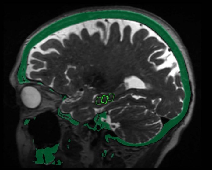
In the study, the delivered focused ultrasound to specific sites in the brain critical to memory in three women, ages 61, 72 and 73, with early-stage Alzheimer’s disease and evidence of amyloid plaques—abnormal clumps of protein in the brain that are linked with Alzheimer’s disease.
The patients received three successive treatments at two-week intervals. Researchers tracked them for bleeding, infection, and edema, or fluid buildup.
Post-treatment brain MRI confirmed that the blood-brain barrier opened within the target areas immediately after treatment. Closure of the barrier was observed at each target within 24 hours.
The team says they were able to open the blood-brain barrier in a very precise manner and document closure of the barrier within 24 hours.
The technique was reproduced successfully in the patients, with no adverse effects.
MRI-guided focused ultrasound involves the placement of a helmet over the patient’s head after they are positioned in the MRI scanner.
The helmet is equipped with more than 1,000 separate ultrasound transducers angled in different orientations.
Each transducer delivers sound waves targeted to a specific area of the brain.
Patients also receive an injection of contrast agent made up of microscopic bubbles. Once ultrasound is applied to the target area, the bubbles oscillate or change size and shape.
While the research so far has focused on the technique’s safety, in the future the researchers intend to study the treatment effects.
One author of the study is Rashi Mehta, M.D., an associate professor.
The study was presented at the annual meeting of the Radiological Society of North America (RSNA).
Copyright © 2019 Knowridge Science Report. All rights reserved.


