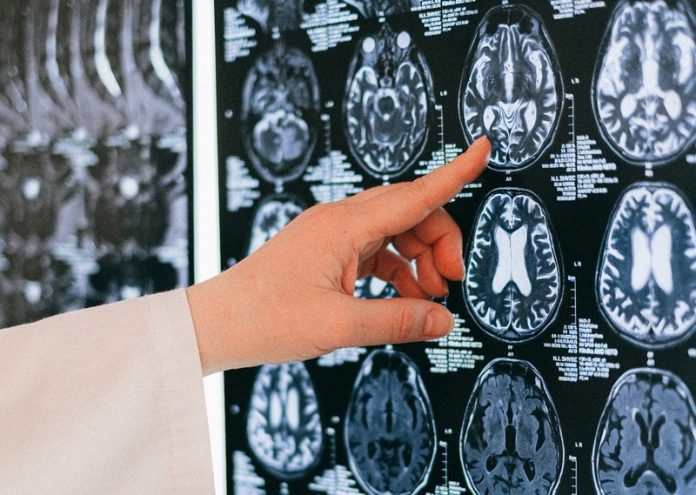
Alzheimer’s disease usually is diagnosed based on symptoms, such as when a person shows signs of memory loss and difficulty thinking.
Up until now, MRI brain scans haven’t proven useful for early diagnosis in clinical practice.
In a new study from Washington University in St. Louis, researchers found that a mathematical analysis of data obtained with a novel MRI approach can identify brain cell damage in people at early stages of Alzheimer’s, before tissue shrinkage is visible on traditional MRI scans and before cognitive symptoms arise.
The technique takes only six minutes to acquire data and can be implemented on MRI scanners that are already used worldwide for patient diagnostics and clinical trials.
It could be a new way to use MRI to diagnose people with Alzheimer’s before they develop symptoms.
In the study, the team tested 70 people ages 60 to 90. Participants completed extensive clinical and cognitive testing to assess their level of cognitive impairment.
The participant group included people with no cognitive impairment as well as those with very mild, mild, or moderate impairments.
Each participant underwent either a PET brain scan or a spinal tap to gauge the number of amyloid plaques in his or her brain. They also underwent MRI brain scans.
The study relies on a new quantitative Gradient Echo (qGRE) MRI technique to show brain areas that are no longer functioning due to a loss of healthy neurons.
While traditional MRI is capable of showing where damaged areas of the brain have decreased in volume, the qGRE technique goes a step further, detecting the loss of neurons that precedes brain shrinkage and cognitive decline.
The researchers applied the qGRE MRI technique to scan the hippocampus, the brain’s memory center, and one of the earliest affected brain regions in Alzheimer’s.
They found the hippocampus often contained a viable tissue section with relatively preserved neurons and a dark matter dead zone virtually devoid of healthy neurons.
These dark matter areas were present in people who tested positive for amyloid but were not yet experiencing symptoms, and they grew larger as the disease progressed.
Compared with traditional MRI measures of brain atrophy, biomarkers for dark matter correlated much better with individual cognitive scores for very mild to moderate dementia.
While each testing approach has its own strengths and weaknesses, the qGRE MRI technique may be poised for early adoption since it is based on MRI technology that is widely available worldwide, is noninvasive, and can be carried out without the use of radioactive tracers.
If you care about Alzheimer’s disease, please read studies that COVID-19 and Alzheimer’s disease are connected, and findings of new method that can predict Alzheimer’s disease with nearly 100% accuracy.
For more information about Alzheimer’s disease, please see recent studies that new non-drug treatment may help prevent Alzheimer’s effectively, and results showing this new method shows great potential for treating Alzheimer’s disease.
The study is published in the Journal of Alzheimer’s Disease and was conducted by Dmitriy Yablonskiy et al.
Copyright © 2022 Knowridge Science Report. All rights reserved.


