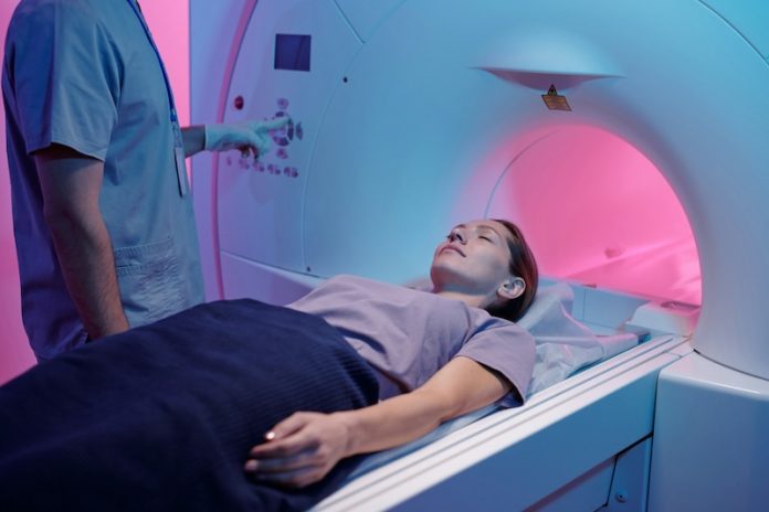
Artificial intelligence (AI) is becoming an essential tool in medical imaging, helping radiologists analyze scans faster and more accurately. However, AI models need a massive amount of labeled data to work well, which means radiologists still spend a lot of time marking images manually.
A new international project led by researchers at Johns Hopkins University offers a solution—AbdomenAtlas, the largest abdominal CT scan dataset ever created.
The AbdomenAtlas dataset includes over 45,000 3D CT scans from 145 hospitals worldwide. It maps 142 different anatomical structures, making it 36 times larger than the next biggest dataset, TotalSegmentator V2.
This massive collection of scans is expected to greatly improve AI training, allowing machines to better detect organs, tumors, and diseases. The dataset and its applications were recently published in the journal Medical Image Analysis.
In the past, radiologists manually labeled organs in CT scans, a process that took thousands of hours. To put the challenge into perspective, marking 45,000 scans with six million anatomical structures would have required an expert to start in 420 BCE—the time of Hippocrates—and work nonstop until 2025. Clearly, this task was too large for humans alone.
To speed up the process, the Johns Hopkins team, led by Professor Alan Yuille, used AI models to handle most of the labeling work. Their approach combined three AI systems trained on existing labeled datasets to predict the anatomy in new, unlabeled scans.
These AI models then highlighted areas that needed review using color-coded attention maps. A team of 12 expert radiologists and medical trainees manually checked and refined the AI’s predictions.
This AI-assisted method dramatically reduced the time needed to annotate scans. For labeling tumors, the process became 10 times faster, while for organs, it was 500 times faster than manual annotation alone.
This allowed the researchers to build the largest fully annotated abdominal organ dataset in less than two years—a task that would have otherwise taken humans more than 2,000 years.
The dataset continues to grow as more scans are added, along with artificial and real tumor samples. These additions will help AI models improve in detecting cancer, diagnosing diseases, and even creating “digital twins” of real patients for personalized medicine.
Beyond training AI, AbdomenAtlas serves as a benchmark for evaluating other medical imaging algorithms. By testing AI models on such a large dataset, researchers can better ensure that these models will work reliably in real-world medical settings.
The team has already used the dataset in competitions, such as the BodyMaps Challenge at an international medical imaging conference, to encourage AI development that is both accurate and practical for clinical use.
Despite its scale, AbdomenAtlas represents only 0.05% of the CT scans taken in the U.S. each year. The researchers hope more hospitals and institutions will collaborate to expand the dataset. They believe that sharing data and working across institutions is essential for advancing AI in medicine.
By creating AbdomenAtlas, the team has provided a powerful resource that could transform medical imaging. As AI continues to evolve, datasets like this will help ensure that machines can assist doctors in diagnosing diseases faster and more accurately than ever before.
If you care about cancer, please read studies that low-carb diet could increase overall cancer risk, and new way to increase the longevity of cancer survivors.
For more information about cancer, please see recent studies about how to fight cancer with these anti-cancer superfoods, and results showing daily vitamin D3 supplementation may reduce cancer death risk.
The research findings can be found in Medical Image Analysis.
Copyright © 2025 Knowridge Science Report. All rights reserved.


