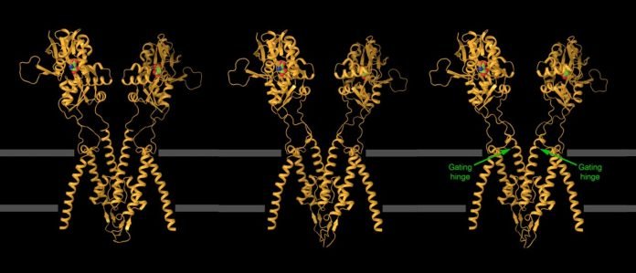
Scientists have recently captured groundbreaking images of one of the brain’s fastest-acting proteins, known as the kainate receptor.
These images are providing crucial clues that could help develop new treatments for epilepsy and other brain disorders.
The findings were published in the journal Nature.
The kainate receptor is vital for communication between neurons in the brain. It works by opening its ion channel quickly and then closing it within milliseconds. This rapid action has made it incredibly difficult for scientists to capture images of the receptor in action.
Alexander Sobolevsky, an associate professor at Columbia University, led the team that succeeded in capturing these images. He explained that the main challenge is the speed at which the receptor works. Freezing the receptor in its active state takes about 30 seconds, but the receptor completes its action in just a few milliseconds.
Despite numerous attempts, scientists had struggled to get clear images of the kainate receptor. This receptor is involved in epilepsy and many other brain disorders, including depression, anxiety, and autism. Because of this, it’s a key target for drug developers. However, only a few approved drugs can safely target the receptor.
To overcome this challenge, Sobolevsky’s team used two innovative methods. First, they found molecules that could stick to the receptor and keep it open longer. They discovered two such molecules: BPAM and lectin Concanavalin A. BPAM could keep the channel open for a few hundred milliseconds, but this wasn’t enough. When they combined BPAM with lectin, the channel stayed open for several seconds.
Maria Yelshanskaya, an expert in electrophysiology, used special recordings to determine the right timing. She found that BPAM or lectin alone couldn’t keep the channel open long enough. However, when used together, they produced a “Eureka moment” for the team by keeping the channel open for several seconds.
Even with this improvement, the conventional method of freezing, which takes about 30 seconds, was still too slow. So, Kirill Nadezhdin, a postdoctoral researcher, designed a robotic plunger that could freeze the sample in less than 3 seconds. This allowed the team to capture the receptor in its open state.
With these two innovations—the molecular stabilizers and the robotic plunger—the team was able to capture detailed images of the kainate receptor in various open positions.
These images are incredibly valuable for drug developers. Some drugs work by blocking ion channels, similar to putting a cork in a bottle. To create these drugs, scientists need precise images of the open channel to ensure a perfect fit.
The images also show how the epilepsy drug perampanel can be optimized to target specific versions of kainate receptors, potentially leading to more precise treatments for patients. Other researchers are already using these images to explore how to improve existing drugs.
The methods developed by Sobolevsky’s team are expected to help other scientists capture more images of fast-acting molecules like the kainate receptor. The more images scientists have, the better their models will be, which could lead to the development of better drugs.
In summary, these groundbreaking images of the kainate receptor provide critical insights that could lead to the development of new, targeted therapies for epilepsy and other brain disorders.
If you care about brain health, please read studies about inflammation that may actually slow down cognitive decline in older people, and low vitamin D may speed up cognitive decline.
For more information about brain health, please see recent studies about common exercises that could protect against cognitive decline, and results showing that this MIND diet may protect your cognitive function, prevent dementia.


