Tiny particles of gold, just a billionth of a meter in size, have been known to kill cancer cells for a long time.
It was thought that the smaller the gold nanoparticles, the faster they would kill cancer cells because they could easily enter the cells and disrupt their functions.
However, recent research has shown that this explanation is too simple.
Scientists at the Institute of Nuclear Physics of the Polish Academy of Sciences (IFJ PAN) in Cracow have discovered a more complex story using a new microscopic technique.
Supported by theoretical analysis from the University of Rzeszow (UR) and Rzeszow University of Technology, they found that the toxicity of gold nanoparticles to cancer cells depends on more than just their size.
Dr. Joanna Depciuch-Czarny from IFJ PAN explains, “We have a top-notch medical and accelerator center for proton radiotherapy.
When reports suggested that gold nanoparticles could enhance this therapy, we decided to make and test them ourselves. We quickly found that their toxicity was not always as expected.”
Gold nanoparticles can be made in various shapes and sizes. Initially, the scientists expected that smaller particles would be more toxic.
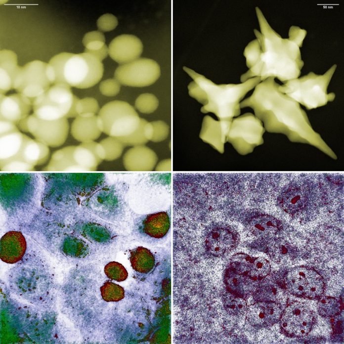
Surprisingly, they found that small spherical nanoparticles of about 10 nanometers were almost harmless to the glioma cells they studied. In contrast, larger star-shaped nanoparticles of about 200 nanometers caused high mortality in the cells.
To understand this contradiction, the scientists used the first holotomographic microscope in Poland. Similar to a CT scanner that uses X-rays to create 3D images of the human body, the holotomographic microscope uses electromagnetic radiation to scan cells without disturbing them.
This technique allows scientists to see a detailed 3D image of the cells and their interiors.
Dr. Depciuch-Czarny says, “Holotomography doesn’t require any sample preparation or introduction of foreign substances into the cells.
We could observe the interactions of gold nanoparticles with cancer cells directly in the incubator, where they were cultured, in real time and with nanometric resolution.”
Using holotomography, the scientists found that while small spherical nanoparticles easily entered the cancer cells, the cells managed to push them out and even began to divide again after initial stress.
In contrast, the large star-shaped nanoparticles punctured the cell membranes with their sharp tips, causing oxidative stress inside the cells. This stress eventually led to apoptosis, or programmed cell death, when the cells could no longer repair the damage.
The research team used data from their experiments to create a theoretical model of how nanoparticles interact with cancer cells.
Dr. Pawel Jakubczyk, a professor at UR, explains, “We developed a differential equation that describes how nanoparticles are taken up by cancer cells over time. Any scientist can use our model to quickly determine the best shape and size of nanoparticles for their research.”
This model can save time and money by reducing the number of experiments needed. It typically takes about two weeks to culture a cell line, but the model can help scientists focus on the most promising nanoparticles.
Additionally, it can help design better-targeted therapies where nanoparticles are absorbed by cancer cells but remain non-toxic to healthy cells.
The Cracow-Rzeszow group is already planning further research. They aim to include more parameters in their model, such as the chemical composition of the nanoparticles and different types of tumors.
They also plan to optimize the efficacy of photo- or proton therapy using various combinations of nanoparticles and tumors.
In summary, gold nanoparticles offer a promising new way to fight cancer. By understanding how these tiny particles interact with cancer cells, scientists can develop more effective and targeted treatments, potentially saving many lives in the future.


