Doctors often face challenges when trying to see deep inside the human body to detect and treat diseases.
The body is a complex network of tiny, winding passageways that can be difficult to access, especially in areas like the cardiovascular and respiratory systems.
Endoscopes, thin flexible tubes with lights and sometimes cameras, are commonly used by doctors to explore these areas.
However, most endoscopes are still too large to reach the body’s smallest spaces, like narrow blood vessels or tiny lung branches. Now, a new type of lens system for endoscopes might change that.
A team led by Professor Arka Majumdar from the University of Washington has designed a special lens system that could allow doctors to see into these tiny, hard-to-reach spaces.
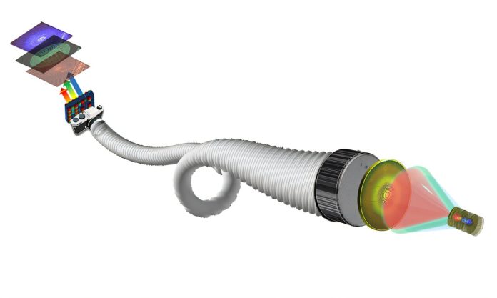
The research, published in Light: Science & Applications, presents a “metalens” – a flat, lightweight lens created from microscopic structures that control light precisely.
This metalens could make endoscopes much smaller and more flexible, allowing doctors to reach areas inside the body that were previously inaccessible.
This new lens system could shrink the size of some endoscopes by more than 50%, making it possible to explore narrow spaces, such as small arteries in the brain or deep blood vessels in the heart.
For patients with cardiovascular issues, this could improve early detection and treatment of serious conditions like heart attacks and strokes, which are leading causes of death worldwide.
Because it uses a real-time camera, the system could give doctors high-quality visual feedback immediately during procedures, helping to reduce medical errors and improve treatment success rates. Importantly, this new metalens system also provides clearer and more detailed images than X-rays without exposing patients to harmful radiation.
The lens works by taking advantage of something called chromatic aberration, a common optical effect where light colors don’t all focus in the same place.
While chromatic aberration can often distort images, Majumdar’s team turned it into a feature: different colors are used to create a three-dimensional, color-coded depth map of the area being viewed. This approach also uses “quantitative phase imaging,” which lets the lens capture a full-color, 3D video in real-time.
Because it doesn’t require complex computing, it’s especially useful for medical procedures where quick feedback is essential.
The tiny metalens is only about 0.5 millimeters wide, roughly the width of five human hairs. Unlike many current endoscopic technologies, this small size and advanced imaging technique allow the device to access small, hard-to-reach parts of the body without needing large equipment.
Now that the research team has demonstrated this new lens concept, they plan to build a prototype to test in models of human organs over the next two years. If successful, they will move forward with clinical studies, with hopes of eventually bringing this technology to the medical market.
The long-term goal is to develop a product that could be widely used in healthcare settings, but the researchers caution that it could take many years to see its full impact.
Professor Eric Seibel, who has spent decades developing endoscopes, sees this as a major step forward. “We’re trying to extend the surgeon’s eyes deeper into the body,” he said.
Though this technology may take time to reach everyday use, it has the potential to greatly improve how doctors examine and treat diseases, making a lasting difference in patient care.
If you care about health, please read studies that vitamin D can help reduce inflammation, and vitamin K could lower your heart disease risk by a third.
For more health information, please see recent studies about new way to halt excessive inflammation, and results showing foods that could cause inflammation.
Source: KSR.


