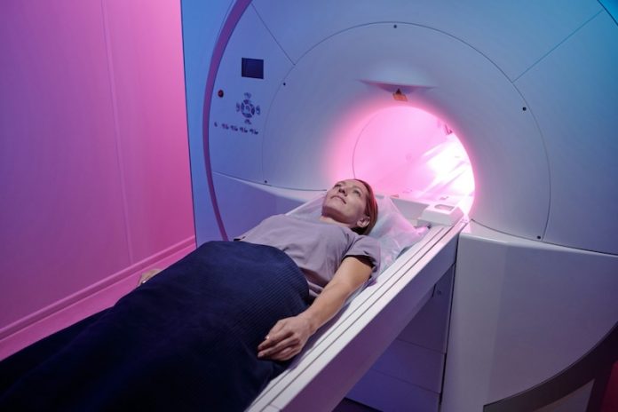
There is a growing interest in combining artificial intelligence (AI) with medical imaging to improve how doctors diagnose and monitor brain diseases.
One exciting area of research focuses on using machine learning to enhance images produced by magnetic resonance imaging (MRI).
Recent studies suggest that ultra-high-field MRI, using a 7 Tesla (7T) system, can provide much sharper and more detailed images than the more common 3T MRI, particularly when examining the brain.
In most hospitals and clinics in the U.S., MRI machines operate at either 1.5T or 3T, with the 3T system being the more powerful option for routine scans.
However, as of 2022, there were only about 100 7T MRI machines being used for diagnostics worldwide, according to the National Institutes of Health.
The limited availability of these advanced machines means that many patients don’t benefit from the higher-resolution images they can provide.
Researchers from the University of California, San Francisco (UCSF), have developed a machine learning algorithm that addresses this issue by creating synthetic 7T images from standard 3T MRI scans.
This algorithm enhances the resolution of 3T images, making them more comparable to the ultra-high-quality 7T images.
The new approach could provide doctors with clearer images of the brain, especially when diagnosing conditions that involve subtle changes in brain tissue, such as traumatic brain injury (TBI) and multiple sclerosis.
The study, which was presented on October 7 at the 27th International Conference on Medical Image Computing and Computer Assisted Intervention (MICCAI), showcases how machine learning can create these high-quality images.
“Our paper introduces a machine-learning model to synthesize high-quality MRIs from lower-quality images,” explained Dr. Reza Abbasi-Asl, an assistant professor of neurology at UCSF and the senior author of the study.
“We demonstrate how this AI system improves the visualization and identification of brain abnormalities captured by MRIs in Traumatic Brain Injury.”
By developing this model, the researchers are taking a significant step toward improving the accuracy and usefulness of MRI scans in clinical settings.
The enhanced images make it easier to see abnormalities in the brain, which could lead to better diagnoses and treatment plans for patients with brain injuries or degenerative diseases.
In the study, the UCSF team collected MRI data from patients diagnosed with mild traumatic brain injury (TBI). They then trained three different neural network models to enhance the standard 3T MRI images.
These AI models were designed to sharpen the details of the images, creating what the researchers call synthetic 7T MRIs. The goal was to make the enhanced 3T images look and function more like those produced by a real 7T machine.
The results were impressive. The new models provided images with enhanced details of the brain tissue affected by mild TBI. For example, the researchers focused on areas of the brain with white matter lesions and tiny bleeds, known as microbleeds, that are difficult to see on regular MRI scans.
In the synthetic 7T images, these lesions and microbleeds appeared clearer and more distinct, allowing for better identification of the problem areas.
The sharper contours of the brain tissue and the clearer separation of adjacent lesions made it easier to analyze the damage caused by the brain injury.
This new approach not only helps with TBI diagnosis but also holds potential for other brain disorders, like multiple sclerosis. In patients with multiple sclerosis, small changes in brain tissue can indicate disease progression.
The enhanced images created by the AI model could make it easier to detect these changes, leading to more accurate diagnoses and improved treatment strategies.
Although the initial results are promising, the researchers acknowledge that more work needs to be done before this technology can be widely used in clinical practice.
They emphasize the importance of further testing to validate the accuracy and reliability of the AI-generated images. Future studies will need to assess the model’s performance in real-world clinical settings and determine how well it holds up under different conditions.
In conclusion, the combination of AI and MRI technology represents a significant advance in medical imaging, offering the possibility of clearer, more detailed brain scans without the need for ultra-high-field MRI machines.
This could help doctors better diagnose and treat patients with brain disorders, improving outcomes for those suffering from traumatic brain injuries, multiple sclerosis, and other conditions that affect the brain.
If you care about brain health, please read studies about how the Mediterranean diet could protect your brain health, and blueberry supplements may prevent cognitive decline.
For more information about brain health, please see recent studies about antioxidants that could help reduce dementia risk, and Coconut oil could help improve cognitive function in Alzheimer’s.
The research findings can be found in Medical Image Computing and Computer Assisted Intervention – MICCAI 2024.
Copyright © 2024 Knowridge Science Report. All rights reserved.


