In a new study, researchers did one of the largest, most comprehensive analysis of autopsies of COVID-19 victims to date.
They found many complex new details about the disease.
The research was conducted by a team at The Mount Sinai Hospital.
An essential contribution of pathology is the understanding of the biology of the disease and the range of organ damage, and for this reason, the team decided to uncompromisingly perform as many autopsies as possible.
Post-mortem examinations (autopsies) are the gold standard for the elucidation of the underlying pathophysiology of the disease.
Despite a rapidly growing body of literature focusing on the clinical impact and molecular microbiology of SARS-CoV-2, autopsy studies have comparatively been few and far between.
To date, the team has performed more than 90 autopsies on deceased COVID-19 patients at Mount Sinai Hospital. The published work analyzes the first 67.
Gross anatomical findings were combined with the clinical history and laboratory data for all 67 patients.
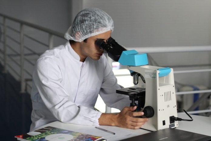
Microscopic examinations were carried out by the team, using special stains, immunochemistry, electron microscopy, and molecular pathology assays.
COVID-19 was initially conceptualized as a primarily respiratory illness, but the Mount Sinai analysis laid out in detail that it also causes damage to the thin layer of cells that line blood vessels (endothelium), which underlies the clotting abnormalities and hypoxia observed in severely ill patients who develop multi-organ failure that leads to death in some patients.
The lungs in nearly all cases showed diffuse damage to the alveoli, the small sacs where oxygen and carbon dioxide are exchanged with the blood.
This damage is the typical microscopic evidence of clinical acute respiratory distress syndrome (ARDS), with most cases showing fibrin (a fibrous, non-globular protein involved in the clotting of blood) and/or platelet thrombi, or clots, to varying extents.
This same pathology is found in most cases of ARDS, including those related to other coronaviruses.
However, the totality of findings in the autopsy series as a whole, with blood clots in multiple other organ systems—most notably the brain, kidney, and liver—reflects endothelial damage as an underlying process, which would also correlate with the activation of the coagulation cascade and persistent elevation of blood markers of inflammation.
The examined brains showed a surprising scarcity of inflammation, with only a few cases showing small foci of chronic inflammation.
However, a surprising number of cases showed microthrombi with small and patchy evidence of tissue death caused by blockage of blood vessels in both peripheral and deep parts of the brain.
These small microinfarcts may explain some of the psychological changes seen in some COVID-19 positive patients.
This study brings new light into the pathophysiology of COVID-19, offering justification for novel treatment plans.
One author of the study is Carlos Cordon-Cardo, MD, Ph.D.
The study was released on the preprint server MedRxiv.
Copyright © 2020 Knowridge Science Report. All rights reserved.


