Scientists at the Allen Institute have made groundbreaking discoveries about how the brain changes as it ages.
By studying mice, they identified specific brain cell types that undergo major changes, especially in a region called the hypothalamus.
This research, published in Nature, may lead to new ways to slow or manage brain aging and reduce the risk of age-related brain disorders.
What changes as the brain ages?
The study revealed that certain brain cells, mostly glial cells, experience significant changes in gene activity with age. Glial cells are support cells in the brain, and the most affected types include:
- Microglia and border-associated macrophages (immune cells in the brain)
- Oligodendrocytes (cells that produce myelin, which protects neurons)
- Tanycytes and ependymal cells (cells involved in metabolism and nutrient regulation)
The researchers found two major shifts in gene activity in aging brains:
- Increased inflammation: Genes linked to inflammation became more active.
- Decreased neuronal function: Genes related to brain structure and function became less active.
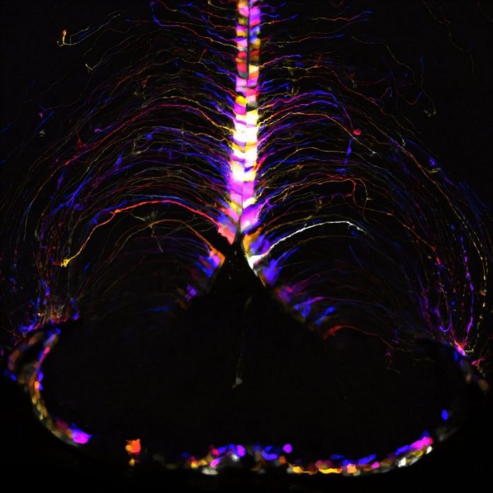
These changes were especially concentrated in the hypothalamus, a small but vital part of the brain that controls metabolism, food intake, and energy balance.
The most significant changes occurred near the third ventricle of the hypothalamus, a region with cells responsible for important processes like regulating diet and metabolism. This “hot spot” may explain how brain aging is linked to lifestyle factors like diet and could influence susceptibility to brain disorders such as Alzheimer’s disease.
“Our hypothesis is that these cell types lose efficiency in processing signals from the environment or diet, which contributes to aging in the brain and body,” explained Dr. Kelly Jin, the study’s lead author.
Using advanced brain-mapping tools and single-cell RNA sequencing, researchers analyzed over 1.2 million brain cells from young (2-month-old) and aged (18-month-old) mice. The aged mice are equivalent to late middle-aged humans. Mice and human brains share many similarities, making this study relevant to understanding human aging.
This detailed mapping allowed scientists to see which cells are most affected by aging, providing valuable insights for future therapies.
Understanding how aging affects specific brain cells opens the door to new treatments. Researchers hope to develop tools to improve the function of these key cells, potentially delaying the aging process and reducing the risk of neurodegenerative diseases.
“This study provides a detailed map of aging in the brain and highlights key players that can be targeted for future therapies,” said Dr. Richard Hodes, director of the National Institute on Aging.
The findings also align with past research suggesting that lifestyle factors, such as diet or intermittent fasting, may influence brain health and longevity.
The study is an important step toward understanding the complexities of brain aging. It sets the stage for developing targeted therapies, including potential drugs or dietary strategies, to preserve brain function into old age.
“This is a beautiful example of why studying specific brain cell types is so important,” said Dr. Hongkui Zeng, a senior researcher at the Allen Institute. “By focusing on the right players, we can uncover changes that would otherwise be hidden.”
As scientists continue to piece together the puzzle of brain aging, these findings bring us closer to strategies that could improve brain health and quality of life as we age.


