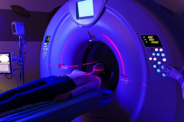
Magnetic resonance imaging (MRI) is a powerful, non-invasive tool for examining the human brain. It uses magnetic fields and radio waves to produce detailed images of soft tissues, making it invaluable for diagnosing brain disorders and conducting neurological research.
However, despite its advantages, MRI has challenges, particularly when patient movement causes blurry images or artifacts, or when variability between scanners affects image consistency.
To address these issues, researchers in Li Wang’s lab at the University of North Carolina have developed two advanced artificial intelligence (AI) models.
These models aim to enhance MRI imaging quality, improve diagnostic accuracy, and provide deeper insights into brain development and aging. Their findings, published in Nature Biomedical Engineering, showcase the potential of AI to transform neuroimaging.
Enhancing Brain Imaging with AI
The first AI model developed by Wang’s team focuses on improving a process called “skull-stripping.” This step involves removing the skull and other non-brain tissues from MRI images to provide a clear view of brain structures.
While essential, skull-stripping often struggles with accuracy, especially when dealing with changes in brain size, tissue contrast, or structural variations across different ages.
Wang’s AI-powered skull-stripping model overcomes these challenges by accurately isolating the brain from surrounding tissues. The model was trained on a large dataset of 21,334 MRI scans collected from 18 sites using diverse imaging protocols and scanners.
By leveraging this broad dataset, the AI model can reliably track brain development and aging across a person’s lifespan. This capability is particularly valuable for understanding how the brain changes over time and for diagnosing conditions linked to these changes.
Improving Imaging Quality with BME-X
The second AI model, known as Brain MRI Enhancement foundation (BME-X), is designed to improve the overall quality of MRI images. MRI scans often vary in resolution, noise levels, and clarity due to differences in scanner models, imaging parameters, and patient movement.
BME-X addresses these issues by enhancing image resolution, reducing noise, correcting motion artifacts, and even harmonizing data from different MRI scanners.
For instance, MRI scanners from manufacturers like Siemens, GE, and Philips each use different imaging protocols, leading to inconsistencies in image quality.
BME-X can process these diverse inputs and produce “harmonized” images, ensuring consistent results regardless of the scanner type. This harmonization is crucial for multi-center studies and clinical trials where consistent imaging data is essential for accurate analysis.
Real-World Applications and Benefits
Both AI models have been rigorously tested on large and diverse datasets. The skull-stripping model was validated on over 21,000 MRI scans, while BME-X was tested on more than 13,000 images from various patient populations and scanner types. In both cases, the models outperformed existing methods in accuracy, quality enhancement, and adaptability.
These advancements have significant implications for clinical care and research. By producing clearer and more reliable images, the AI models can improve early detection, diagnosis, and monitoring of neurological conditions.
They can also facilitate studies that involve multiple research centers or imaging modalities, such as CT scans, by standardizing imaging protocols and reducing variability.
For example, the ability of BME-X to correct motion artifacts and enhance low-resolution images could make it easier to work with younger patients, who often have difficulty staying still during scans. Similarly, harmonized data can improve collaborations between institutions and lead to more robust research findings.
A Step Toward Standardized Imaging
The work from Wang’s lab highlights the transformative potential of AI in neuroimaging. By addressing long-standing challenges in MRI technology, these models pave the way for better diagnostics, more effective treatments, and deeper insights into brain health.
Additionally, the adaptability of these models to other imaging techniques, like CT scans, suggests their benefits could extend beyond MRI.
As imaging technologies continue to evolve, these AI models represent a significant step toward creating standardized and reliable imaging protocols, ultimately improving outcomes for patients and advancing the field of neuroscience.
If you care about brain health, please read studies about vitamin D deficiency linked to Alzheimer’s and vascular dementia, and blood pressure problem at night may increase Alzheimer’s risk.
For more information about brain health, please see recent studies about antioxidants that could help reduce dementia risk, and epilepsy drug may help treat Alzheimer’s disease.
The research findings can be found in Nature Biomedical Engineering.
Copyright © 2025 Knowridge Science Report. All rights reserved.


