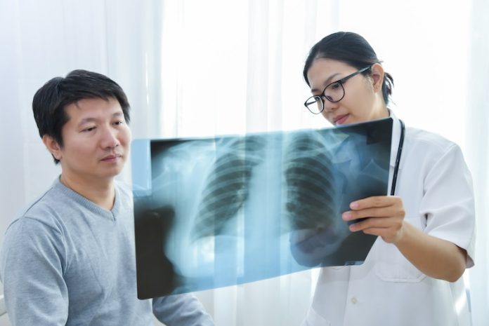
Scientists at Newcastle University in the UK have developed a groundbreaking scanning technique that shows how lungs work during breathing.
This new method allows doctors to see the effects of treatments on lung function as they happen and could help identify problems earlier, especially in patients with asthma, chronic obstructive pulmonary disease (COPD), or those who have had lung transplants.
The technique uses a harmless gas called perfluoropropane, which is easy for patients to inhale and exhale.
Once the gas is breathed in, doctors use an MRI scanner to capture detailed images of the lungs, showing where the gas travels. These scans reveal which parts of the lungs are functioning well and which are not.
Professor Pete Thelwall, an expert in magnetic resonance physics at Newcastle University, led the research.
He explained, “Our scans highlight areas of the lungs that aren’t working properly, and we can see how treatments improve airflow. For instance, when a patient uses asthma medication, we can watch in real time how their lungs respond and improve.”
This method goes beyond traditional lung tests by providing a clear picture of how air flows through different parts of the lungs. Doctors can measure which areas are well-ventilated and which have blockages or damage, helping them understand the severity of a patient’s lung condition.
The research team tested the method on patients with asthma and COPD. In one study, published in the journal Radiology, they used the scanning technique to evaluate the effects of a common asthma medication, salbutamol.
The scans showed exactly how much the medication improved airflow in the lungs. This approach could be especially useful in testing new treatments for lung diseases.
Another important application of this technology is in lung transplant patients. These individuals often face complications, such as their immune system rejecting the donor lungs.
In a separate study published in JHLT Open, the team scanned patients who had received lung transplants. They compared patients with healthy lung function to those experiencing chronic rejection, a common problem where the immune system attacks the small airways of the donor lungs.
The scans showed that in patients with chronic rejection, air struggled to reach the outer parts of the lungs, indicating damage to the tiny airways. This type of damage is not always easy to detect with traditional tests, but the new scanning method revealed it clearly.
Professor Andrew Fisher, a specialist in respiratory transplant medicine at Newcastle University and co-author of the study, explained the potential impact: “This new scan may allow us to detect changes in transplanted lungs earlier, even before traditional lung tests show any problems.
By catching these issues sooner, we could start treatment earlier and help protect the lungs from further damage.”
The researchers believe this technique could improve care for lung transplant recipients and patients with other lung conditions. By detecting early changes in lung function, doctors could provide more timely and effective treatments, improving outcomes for many people.
This innovative approach not only offers hope for better lung disease management but also sets the stage for more personalized and accurate treatments in the future.
If you care about lungs, please read studies about a review of COPD-friendly foods for lung health, and can Vitamin C and E help fight lung cancer.
For more health information, please see recent studies about how diet influences lung health, and these vegetables could benefit your lung health.
The research findings can be found in Radiology.
Copyright © 2024 Knowridge Science Report. All rights reserved.


