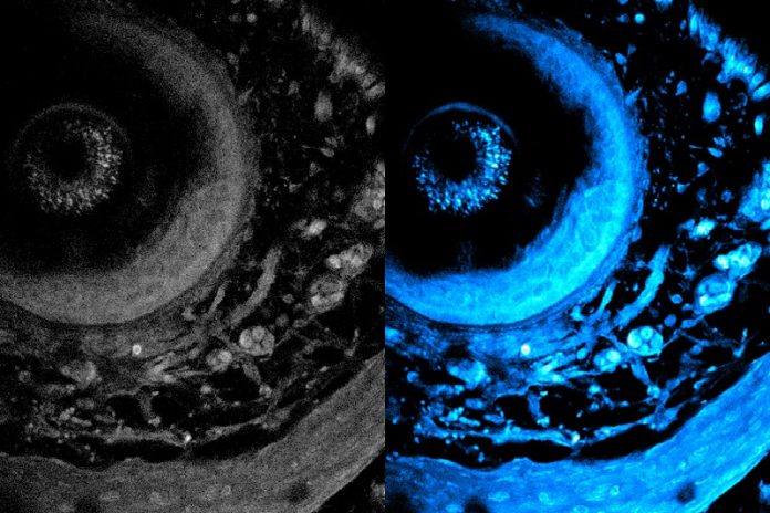
MIT researchers have developed a groundbreaking imaging method that can penetrate twice as deep into living tissue as previous techniques.
This new method allows scientists to capture detailed images of cells without damaging or altering the tissue, opening up exciting possibilities for cancer research, drug discovery, and understanding immune responses.
Traditional imaging methods require tissue to be cut or stained with dyes to reveal details about its structure and function.
While helpful, this process kills the tissue, making it impossible to study dynamic processes in living cells.
Even advanced noninvasive imaging methods are limited by how deeply light can penetrate into tissue.
As light travels through tissue, it scatters, reducing image clarity and limiting how much researchers can see.
The MIT team’s new approach uses specialized lasers and innovative technology to overcome these challenges. Instead of relying on dyes or cutting tissue, they use a laser to illuminate specific molecules naturally present in the tissue. These molecules then emit light, revealing cellular structures and metabolic activity without altering the tissue.
The breakthrough came from developing a “fiber shaper,” a device that bends optical fibers to customize the laser light’s properties. By fine-tuning the light’s color and intensity, the researchers minimized scattering and maximized clarity. This allowed the laser to penetrate deeper into the tissue while maintaining a high-resolution image.
With this technique, the team achieved imaging depths of over 700 micrometers—more than double the previous limit of 200 micrometers. They also improved imaging speed, capturing detailed information about cells and their movements in real time.
“This work shows a significant improvement in how deeply we can see into living tissue,” said Sixian You, an assistant professor at MIT and senior author of the study. “It opens up new possibilities for studying metabolic activity deep within biological systems.”
This method could have transformative impacts on areas like cancer research, neuroscience, and tissue engineering. For example, researchers in MIT’s Roger Kamm and Linda Griffith labs are working on growing organoids—tiny, lab-engineered tissues that mimic the structure and function of organs. Until now, it has been challenging to observe the internal development of organoids without damaging them.
The new imaging technique allows scientists to study organoids noninvasively, monitoring their growth and metabolism over time. It could also help researchers see how cells respond to drugs in real time, improving the process of developing new treatments.
The team is now working to enhance the resolution of their imaging system further and reduce noise to allow even deeper tissue imaging with minimal light exposure. They are also developing algorithms to reconstruct high-resolution 3D images of biological samples.
“By pushing the limits of imaging depth and speed, we’re providing scientists with a powerful tool to explore living tissues in their natural state,” said You. “This could lead to real-world breakthroughs in medicine and biology.”
This research, supported by MIT and several national grants, marks a significant step forward in understanding living systems and solving complex medical problems.


