Researchers from the University Hospital Bonn (UKB) and the University of Bonn have uncovered new insights into how tiny, subtle eye movements help us see sharp details.
Our vision starts with light-sensitive cells in the retina, a layer at the back of our eyes.
A central region in the retina, called the fovea, is responsible for fine, detailed vision.
Packed densely with color-sensitive cells known as cone photoreceptors, the fovea allows us to detect small details, similar to the way pixels work in a camera.
However, unlike camera pixels, the cone cells are not evenly distributed across the retina and vary from person to person.
The researchers used high-tech imaging tools to study how these small eye movements interact with the fovea’s cone cells to optimize sharp vision. Their findings were published in the journal eLife.
Our eyes continuously make small, involuntary movements, even when we fix our gaze on an object.
According to Dr. Wolf Harmening, head of the AOVision Laboratory at UKB, these tiny movements, known as fixational eye movements, help convey fine details of the visual world to the brain.
One particular type of movement, called drift, moves the eye very slightly while we focus on an object. The new study is the first to explore how drift movements and the arrangement of cone cells in the fovea work together to enhance our ability to see fine details.
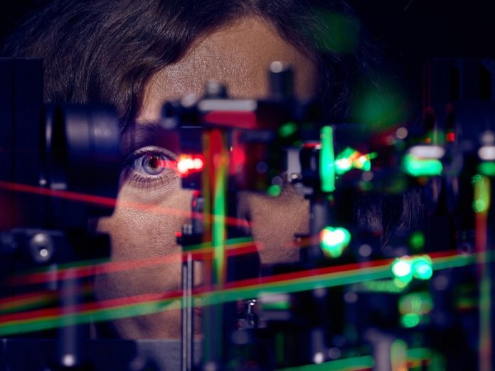
To examine this relationship, the researchers used a special imaging tool called an adaptive optics scanning light ophthalmoscope (AOSLO)—the only one available in Germany. This tool allowed them to see the fovea’s cone cells with high resolution and observe how the eye moves. Sixteen healthy participants were tested on their ability to see tiny details while completing a visual task.
By tracking how visual information was processed by different photoreceptor cells, the researchers were able to analyze each participant’s eye movements and how these movements aligned with the cone cell density in their fovea.
The results showed that people can see even finer details than the number of cone cells in their fovea would suggest. “This suggests that cone cell arrangement only partly explains our visual sharpness,” says Dr. Harmening.
The study also found that drift movements precisely align with areas of higher cone cell density, helping to bring visual stimuli into focus. In just a few milliseconds, the drift movements adjust to focus on areas of the retina with more cone cells, enhancing our sharp vision.
Jenny Witten, a Ph.D. student and the study’s first author, explained that during these subtle eye movements, the retina is guided to the areas with the highest cone density, boosting visual clarity. The length and direction of these movements are essential in keeping vision sharp.
These findings could have important implications. By understanding how the eye naturally moves to achieve sharp vision, scientists may gain insights into eye-related and brain-related conditions that affect vision.
This knowledge could also aid in the development of advanced technologies, like retinal implants, which aim to restore sight for people with visual impairments.
The study highlights how the human eye has evolved to maximize vision through tiny, automatic adjustments, a system that might inspire new ways to enhance or restore vision.
If you care about eye health, please read studies about how vitamin B may help fight vision loss, and MIND diet may reduce risk of vision loss disease.
For more information about eye disease, please see recent studies about how to protect your eyes from glaucoma, and results showing this eye surgery may reduce dementia risk.


