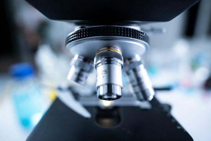
An international team of scientists, led by Trinity College Dublin, has developed an innovative imaging method using advanced microscopes.
This breakthrough significantly reduces the time and radiation needed for imaging, benefiting various fields, including materials science and medicine.
The new method promises to deliver improved imaging for sensitive materials, such as biological tissues, which are especially vulnerable to damage.
Currently, scanning transmission electron microscopes (STEMs) use a highly focused beam of electrons to scan samples, creating images point by point.
At each point, the beam pauses for a set amount of time to gather signals. This process is similar to how cameras use photographic film, with a constant exposure time everywhere.
While effective, this method risks damaging samples because electrons continue to hit the sample until the time for each pixel has passed.
The new method changes this approach by focusing on the fundamental logic of imaging. Instead of observing over a fixed time and counting the detected events (electrons scattered from the sample), the team developed an event-based detection system.
This system measures the varying time it takes to detect a set number of these events.
The new mathematical theory behind this method shows that the first electron detected at each point provides a lot of information, but additional electrons provide diminishing returns.
Since every electron can damage the sample, this method allows for turning off the illumination at the peak of imaging efficiency, using fewer electrons to create high-quality images.
To put this theory into practice, the team has patented a technology called Tempo STEM, developed jointly with IDES Ltd.
This technology uses a high-tech “beam blanker” to shut off the beam once the desired precision at each measurement point is achieved.
Dr. Lewys Jones, the lead researcher from Trinity College Dublin’s School of Physics, explains, “Combining two state-of-the-art technologies in such an exciting way delivers a real leap in the microscope’s capabilities.
Giving microscopists the ability to ‘blank’ or ‘shutter’ the electron beam on and off in nanoseconds in response to real-time events has never been done before.”
This new approach reduces the overall radiation dose needed to produce high-quality images and eliminates excess radiation that only provides diminishing returns.
It also prevents unnecessary damage to the sample.
Dr. Jon Peters, the first author of the research, adds, “We often think of electrons as relatively mild in terms of radiation, but when they are fired at tiny biological samples at speeds of around 75% the speed of light, they can cause significant damage.
This has been a major issue for microscopy, as the images you get back could be unusable or misleading. This is especially problematic when making decisions on future battery materials or catalyst development.”
This breakthrough in microscopy represents a significant advancement in imaging technology, making it possible to study delicate materials more effectively.
By reducing the time and radiation required, the new method promises to deliver better imaging results without damaging sensitive samples, paving the way for future advancements in various scientific fields.


