At the U.S. Department of Energy’s Argonne National Laboratory, scientists have revolutionized X-ray microscopy with a new method that allows them to analyze data as it’s being collected.
This innovative approach, incorporating machine learning, speeds up experiments and enhances data processing by over 100 times while reducing data volume by 25 times.
X-ray microscopes are crucial tools for examining extremely small structures, even those just a few atoms across.
Traditionally, these microscopes generate vast amounts of data that even powerful supercomputers struggle to process efficiently.
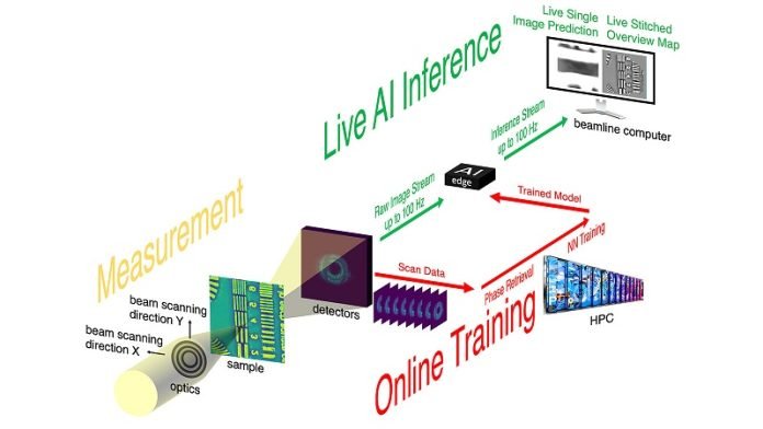
The new technique developed at the Advanced Photon Source (APS), a research facility at Argonne, addresses this challenge by using a neural network—a form of artificial intelligence—to process data on the fly.
Mathew Cherukara, an Argonne group leader and one of the study’s authors, explained that current methods can’t keep up with the massive data flow from these advanced microscopes.
With the old approach, analyzing the data could take days or even weeks without a supercomputer, and hours with one.
The team’s breakthrough involves a technique called streaming ptychography, which works similarly to video streaming services like Netflix.
It analyzes X-ray microscopy data in real time, allowing scientists to immediately adjust the experiment based on the insights gained. This means if researchers spot something intriguing during an experiment, they can explore it right away without waiting for lengthy data analysis.
Antonino Miceli, another Argonne group leader involved in the study, highlighted the benefits of this real-time data processing.
It enables scientists to make quick decisions that could lead to new discoveries, enhancing the efficiency and output of their research.
The neural network essentially learns from the initial data collected during an experiment. As more data is gathered, the network becomes better at predicting and processing new information rapidly.
This setup eliminates the need to send vast amounts of data to remote supercomputers for processing, cutting down on delays.
The streaming ptychography method was tested with high-performance computers at the Argonne Leadership Computing Facility. Initially, these computers performed complex calculations to interpret patterns created by X-ray beams.
This information was then used to train the neural network, which could subsequently carry out similar calculations much faster and at a lower computational cost.
This advancement is not just a leap for X-ray microscopy but could also enhance other types of microscopy, such as electron and optical, by reducing the time and computational power needed for data processing.
The approach also decreases the risk of damaging samples during imaging, making it suitable for delicate materials.
Overall, this new method marks a significant step forward in microscopy, enabling quicker and more efficient experiments and potentially leading to faster scientific discoveries.
A paper detailing these findings was published in Nature Communications.


