Researchers at the Max Planck Institute for Intelligent Systems in Stuttgart have stumbled upon a surprising discovery that could revolutionize medical imaging.
During an experiment initially aimed at drug delivery, they found that nanometer-sized diamond particles could potentially replace gadolinium as a contrast agent in MRI scans.
For over three decades, gadolinium has been a standard contrast agent used to enhance the clarity of MRI images, helping doctors detect tumors, inflammation, and vascular abnormalities.
However, gadolinium has its drawbacks—it tends to accumulate in the brain and kidneys and can linger in the body for months to years, with its long-term effects still unknown.
Moreover, it can leak into surrounding healthy tissue and cause various side effects.
The accidental discovery occurred when Dr. Jelena Lazovic Zinnanti, a scientist at the Max Planck Institute, was conducting an experiment with diamond particles embedded in gelatin capsules.
These capsules were designed to rupture when heated, releasing their contents. Initially, Jelena used gadolinium to track the position of the diamond dust but faced issues with the gadolinium leaking out of the capsules.
Frustrated, Jelena decided to remove gadolinium from her experiments.
To her surprise, when she later took MRI images, the capsules containing only diamond dust still appeared brightly on the scans, showing better signal enhancement than when gadolinium was used.
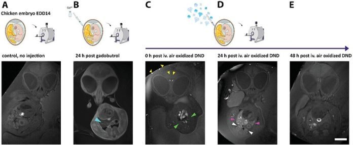
Curious about this phenomenon, Jelena further tested the diamond dust in live chicken embryos.
She discovered that unlike gadolinium, the diamond particles did not diffuse out of the blood vessels but remained contained and continued to provide a bright signal in MRI images days later.
This finding suggested that diamond dust might serve as a more stable and potentially safer contrast agent.
While diamond dust’s ability to enhance MRI signals remains somewhat of a mystery, Jelena hypothesizes that the carbon structure of the diamonds, possibly containing slight magnetic properties due to defects in their crystal lattice, might be responsible for their effectiveness as contrast agents.
The research, now published in the journal Advanced Materials, marks a significant step forward in the search for safer MRI contrast agents. However, more studies are needed to understand fully why diamond dust works so well and to confirm its safety in humans.
If proven safe and effective, diamond dust could one day replace gadolinium, offering a non-toxic alternative that doesn’t accumulate in the body. This would be a major advancement in medical imaging, providing clearer, safer scans and peace of mind for patients undergoing MRI procedures.
Source: Max Planck Society.


