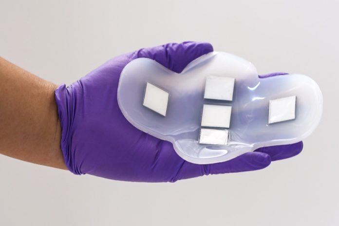
MIT researchers have created an extraordinary ultrasound monitor that can be worn like a patch.
This device makes it easier to image internal organs without needing a professional operator or any messy gel, commonly used in traditional ultrasound procedures.
In a recent study, the MIT team demonstrated that their new patch could effectively image the bladder and measure its fullness.
This development is especially beneficial for people with bladder or kidney disorders, as it allows them to track their organ’s functioning more conveniently.
The researchers believe that this technology can be adapted to monitor other organs just by repositioning the ultrasound array and adjusting the frequency of the signal. This means it could potentially be used for early detection of deep-seated cancers, like ovarian cancer.
Canan Dagdeviren, an associate professor at MIT’s Media Lab and the senior author of the study, emphasized the technology’s versatility. It can identify and characterize various diseases within the body, not just limited to bladder-related conditions.
The idea for this ultrasound patch partly stemmed from a personal experience. Dagdeviren was motivated to work on this project after her brother, diagnosed with kidney cancer, struggled with bladder emptying post-surgery.
She envisioned how such a monitor could aid patients like her brother or those with other kidney or bladder issues.
Currently, measuring bladder volume is only possible with a traditional, bulky ultrasound probe in a medical facility. The team’s goal was to create an at-home wearable alternative. To achieve this, they designed a flexible, silicone rubber patch embedded with five ultrasound arrays made from a newly developed piezoelectric material.
The arrays are arranged in a cross-shape to cover the entire bladder, which measures around 12 by 8 centimeters when full.
The patch’s polymer is naturally adhesive, making it easy to attach and remove. It stays in place when worn under clothing like underwear or leggings.
Collaborating with Massachusetts General Hospital, the researchers conducted a study involving 20 patients with varying body mass indexes (BMIs).
The subjects were imaged with full, partially empty, and completely empty bladders. The patch’s images were comparable in quality to traditional ultrasound and effective across all BMIs.
This patch does not require any gel or pressure application, unlike regular ultrasound probes, as its field of view is large enough to encompass the entire bladder.
The MIT team is currently developing a portable device, similar in size to a smartphone, to view the images. This advancement is part of a larger goal to create a range of devices that can bridge the information gap between clinicians and patients.
Beyond bladder monitoring, the researchers aim to adapt this technology to image other organs like the pancreas, liver, or ovaries. They plan to customize the ultrasound signal’s frequency by developing new piezoelectric materials. For some organs that are deeper within the body, the team is considering implants rather than wearable patches.
This innovation could become a major focus in ultrasound research and future medical device designs. Anantha Chandrakasan, dean of MIT’s School of Engineering and co-author of the study, sees it as a foundation for more collaborations across various scientific fields.
In summary, this new ultrasound patch by MIT researchers offers a simple, non-invasive way for individuals to monitor their bladder and potentially other organs, bringing high-tech medical monitoring into the comfort of one’s home.


