Age-related brain disorders such as Alzheimer’s disease often develop slowly across a lifetime. However, they usually aren’t detected until symptoms have already begun.
This reality has led teams of researchers, led by scientists at Brown University, to explore whether these neurodegenerative diseases could be detected much earlier, perhaps even decades before symptoms appear.
The Potential of Routine Eye Exams
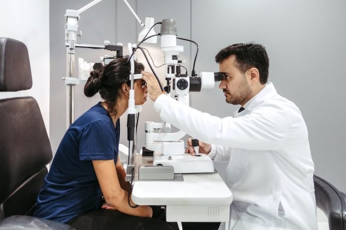
In their new study, these researchers suggest it might be possible to detect these diseases early, through something as simple as a routine eye exam.
They showed in the study, published in Nature Communications, that it is possible to track changes in mice brain blood vessels over a long time.
These blood vessels could provide key clues for the early detection of diseases like Alzheimer’s, Parkinson’s, Huntington’s disease, and multiple sclerosis.
A Closer Look at the Study
The researchers hope to use their method to image the retina in mice, looking for these biomarkers.
From there, they aim to scale up to imaging the retina of humans, tracking how their blood vessels change.
In the study, they used imaging technology to image the brain of the same animal repeatedly for almost one year. This method allowed them to measure the properties of brain blood vessels.
This potentially opens a path to predicting when someone is at risk for developing neurodegenerative diseases, enabling early treatment.
The Role of Cerebral Blood Vessels
Tracking how cerebral blood vessels change over a long time in people who develop age-related neurodegenerative diseases has been a longstanding goal for scientists.
It’s believed that cerebral blood vessels in people who develop brain diseases show signs of degradation and decline decades before symptoms start.
If changes in blood vessels in the brain or retina can be detected over long periods, it may be possible to predict the onset of these diseases.
An Innovative Method
The research team created a novel method that combines advanced imaging techniques and AI algorithms. This method tracks changes in the dynamics and anatomy of brain blood vessels.
They used this method to measure these changes in 25 different mice for more than seven months.
The researchers focused on a non-invasive imaging test called optical coherence tomography (OCT). They adapted multiple OCT techniques to image brain blood vessels.
They then used image processing algorithms to search for patterns in the data they collected.
Through their analysis, they noticed differences between normal age-related changes and changes brought on by the disease.
They found several potential biomarkers, such as large blood vessels getting thinner and blood flow getting lower. Also, the network pattern of vessels changed significantly, compared to normal aging animals.
The Path Forward
The ultimate goal of the researchers is to refine their method and collect enough data so that their algorithms can predict, through regular eye exams, the chance of a person developing neurodegenerative diseases years before they actually do.
While they still have a long way to go to achieve this goal, their proof of concept in mice is a promising start.
They plan to look closer at the biomarkers they found and identify more in the coming years. The next step is to image blood vessels in the retinas of mice.
If you care about health, please read studies about how Mediterranean diet could protect your brain health, and the best time to take vitamins to prevent heart disease.
For more information about health, please see recent studies about plant nutrients that could help reduce high blood pressure, and these antioxidants could help reduce dementia risk.
The study was published in Nature Communications.
Copyright © 2023 Knowridge Science Report. All rights reserved.


