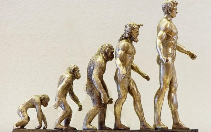
If evolutionary biologist Terence D. Capellini were to rank the body parts that make us quintessentially human, the pelvis would place close to the top.
After all, its design makes it possible for humans to walk upright on two legs (unlike our primate cousins) and it makes it possible for mothers to give birth to babies with large heads (therefore big brains).
On an anatomical level, the pelvis is well understood, but that knowledge starts to break down when it comes to how and when this uber-important structure takes its shape during development.
A new study from Capellini’s lab is changing that. Published in Science Advances, the work shows when during pregnancy the pelvis takes shape and identifies the genes and genetic sequences that orchestrate the process.
The work can one day shed light on the genetic origin of bipedalism and open the door for treatments or predictors of hip joint disorders, like hip dysplasia and hip osteoarthritis.
“This paper is really focused on what all humans share, which are these changes to the pelvis that allowed us to walk on two legs and allowed us to give birth to a large fetal head,” said Capellini, a newly tenured Professor in the Department of Human Evolutionary Biology and senior author on the study.
The study shows that many of the features essential for human walking and birth form around the 6- to 8-week mark during pregnancy. This includes key pelvic features unique to humans, like its curved and basin-like shape.
The formation happens while bones are still cartilage so they can easily, curve, rotate, expand, and grow.
The researchers also saw that as other cartilage in the body begins to turn into bone this developing pelvic section stays as cartilage longer, so it has time to form properly.
“There appears to be a stalling that happens and this stalling allows the cartilage to still grow, which was pretty interesting to find and surprising,” Capellini said.
“I call it a zone of protection.”
The researchers performed RNA sequencing to show which genes in the region are actively triggering the formation of the pelvis and are stalling ossification, which normally turns softer cartilage to hard bone.
They identified hundreds of genes that are turned either on or off during the 6- to 8-week mark to form the ilium in the pelvis, the largest and uppermost bones of the hip with blade-like structures that curve and rotate into a basin to support walking on two legs.
Compared to chimpanzees and gorillas, the shorter and wider reorientation of our pelvic blades make it so humans don’t have to shift the mass of our weight forward and use our knuckles to walk or balance more comfortably.
It also helps increase the size of the birth canal. Apes on the other hand have much narrower birth canals and more elongated ilium bones.
The researchers started the study by comparing these differences in hundreds of skeletal samples of humans, chimpanzees, and gorillas.
The comparisons demonstrated the striking effects that natural selection has had on the human pelvis, the ilium in particular.
To see when the ilium and pelvic elements forming the birth canal began to take shape, the researchers examined 4- to 12-week-old embryos under a microscope with the consent of people who had legally terminated their pregnancies.
The researchers then compared samples from the developing human pelvis’ with mouse models to identify the on and off switches triggering the formation.
The work was led by Mariel Young, a former graduate researcher in Capellini’s lab who graduated in 2021 with her Ph.D. The study was a collaboration between Capellini’s lab and 11 other labs in the U.S. and around the world. Ultimately, the group wants to see what these changes mean for common hip diseases.
“Walking on two legs affected our pelvic shape, which affects our disease risk later,” Capellini said.
“We want to reveal that mechanism. Why does selection on the pelvis affect our later disease risk of the hip, like osteoarthritis or dysplasia. Making those connections at the molecular level will be critical.”


