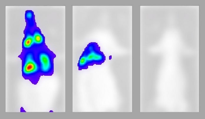
Video images capture for the first time in live animals the inexorable spread of the COVID-19 virus, tracking the infection as it moved from the noses of mice to the lungs and other organs over the course of six days, in a new study led by researchers at Yale and Université de Montréal.
While the images record the sometimes deadly march of SARS-CoV-2, the virus that causes COVID-19, they also show how the introduction of antibodies collected from humans who recovered from the virus can prevent or treat infection.
However, the research also revealed that antibodies lacking the ability to recruit killer immune system cells are less effective at combating infection.
The study, published in the journal Immunity, was led by Priti Kumar, Pradeep Uchil, and Walther Mothes, all from Yale School of Medicine, as well as Andrés Finzi from Université de Montréal.
“For the first time, we were able to visualize the spread of the SARS-CoV-2 in a living animal in real time, and importantly, the sites at which antibodies need to exert effects to halt progression of infection,” said Kumar, associate professor of infectious diseases at Yale School of Medicine and a co-corresponding author of the paper.
For the study, co-lead authors Irfan Ullah, a Yale postdoctoral associate, and Jérémie Prévost, from Montreal, used bioluminescent tagging and advanced microscopy to track the spread of the virus down to the level of single cells.
In mice, the virus took a route that has become familiar to doctors treating human patients, with high viral loads first appearing in nasal passages and then travelling quickly to lungs and eventually other organs.
The mice eventually died when virus reached the brain.
The researchers then used plasma from humans who had recovered from COVID-19 to treat some of the infected mice, which halted the spread of the virus even when administered as late as three days after infection.
When these antibodies were administered prior to infection with the virus, researchers found, they prevented infection altogether.
“The live reporting of virus spread by imaging can be harnessed to rapidly discern whether treatments will work or not in as little as three to five days, a crucial time-saving feature to develop countermeasures for current and future pandemics” said Uchil, a research scientist in Mothes’ lab in the Department of Microbial Pathogenesis at Yale.
Researchers found that not all antibodies worked equally well, however. Antibodies have two main roles.
Neutralizing antibodies bind to and prevent viruses from entering cells.
Then, a second part of the antibody exhibits what are known as “effector” functions, which are necessary to signal the immune system to attack and kill cells that are infected.
“Antibodies are polyfunctional molecules with several properties,” Finzi said.
“In this study we show that their capacity to ‘call for help’ from other cells in the immune system and eliminate infected cells is required to provide optimal protection.”
Added Kumar: “We used to think neutralizing the virus was enough to prevent infection, but antibodies have to be present at the right time in the right place in the body and in right amount.
Without the effector function, the neutralizing activity alone is not as effective.”
Written by Bill Hathaway.


