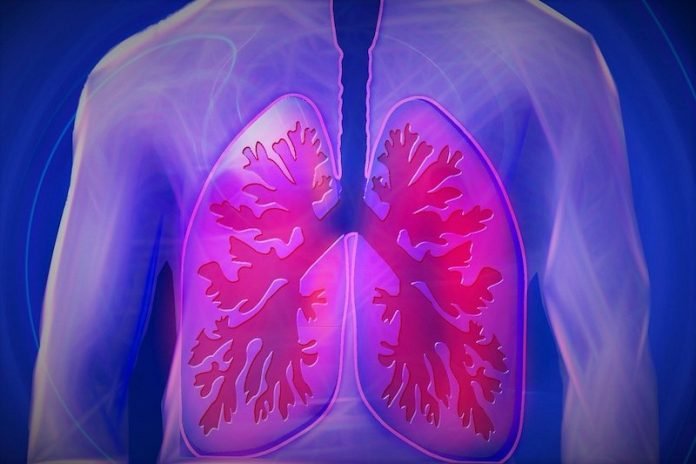
In a new study, researchers found that COVID-19 can cause big dilation of the blood vessels of the lung, specifically the capillaries.
This vasodilation is contributing to the very low oxygen levels seen in COVID-19 respiratory failure and also helps explain why the disease behaves differently than classic acute respiratory distress syndrome (ARDS).
The research was conducted by a team at Mount Sinai.
In classical ARDS, pulmonary inflammation leads to leaky pulmonary blood vessels that flood the lungs with fluid, making the lungs stiff and impairing oxygenation.
Many patients with COVID-19 pneumonia demonstrate severe hypoxemia that is markedly out of proportion to the degree of lung stiffness.
This disconnect between gas exchange and lung mechanics in COVID-19 pneumonia has raised the question of whether the mechanisms of hypoxemia in COVID-19 differ from those in classical ARDS.
The researchers were initially assessing cerebral blood flow in mechanically ventilated COVID-19 patients with altered mental status to look for, among other things, abnormalities consistent with a stroke.
During this study, agitated saline—saline with tiny microbubbles—is injected into the patient’s vein to determine if those microbubbles appear in the blood vessels of the brain.
Under normal circumstances, these microbubbles would travel to the right side of the heart, enter the blood vessels of the lungs, and ultimately get filtered by the pulmonary capillaries, because the diameter of the microbubbles is bigger than the diameter of the pulmonary capillaries.
If the microbubbles are detected in the blood vessels of the brain, it implies that either there is a hole in the heart, so that blood can travel from the right to the left side of the heart without going through the lungs, or that the capillaries in the lungs are abnormally dilated, allowing the microbubbles to pass through.
In the study, 18 mechanically ventilated patients with severe COVID-19 pneumonia were tested.
Fifteen out of the 18 (83 percent) patients had detectable microbubbles, indicating the presence of abnormally dilated pulmonary blood vessels.
In addition, the pulmonary vasodilations may explain the disproportionate hypoxemia seen in many patients with COVID-19 pneumonia.
The findings show that the virus wreaks havoc on the pulmonary vasculature in a variety of ways. This study helps explain the strange phenomenon seen in some COVID-19 patients known as ‘happy hypoxia,” where oxygen levels are very low, but the patients do not appear to be in respiratory distress.
The team says if these findings are confirmed in larger studies, pulmonary microbubble transit may potentially serve as a marker of disease severity or even a surrogate endpoint in therapeutic trials for COVID-19 pneumonia.
One author of the study is Alexandra Reynolds, MD, Assistant Professor of Neurosurgery, and Neurology.
The study is published in the American Journal of Respiratory and Critical Care Medicine.
Copyright © 2020 Knowridge Science Report. All rights reserved.


