In a new study, researchers have developed a new way to detect prostate cancer.
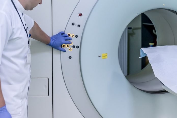
The method includes a biopsy guided by magnetic resonance imaging (MRI). It can be used together with the traditional method to increase the rate of prostate cancer detection.
The research was conducted by a multidisciplinary team of UCLA physicians.
About one million men in the U.S. undergo biopsies to determine whether they have prostate cancer every year.
The biopsy procedure is guided by ultrasound imaging, but this method cannot clearly display the location of tumors in the prostate gland.
Previous research has found that MRI can allow doctors to see specific lesions in the prostate and only take tissue samples from those spots.
But the two sampling methods often aren’t used in combination.
In the 300-person study, 248 men had a prostate lesion visible on MRI. An additional 52 men in the trial had no lesion visible on MRI.
The team combined both sampling methods and found it led to the detection of up to 33% more cancers than standard methods.
The cancer detection rate surpasses that from using either method alone.
The findings suggest that different biopsy methods identify different tumors, and they could help lead to an important change in how prostate biopsies are performed.
The study is the first to directly compare the different biopsy sampling methods in the same group of men.
The team suggests that men being assessed for prostate cancer should first receive an MRI before a biopsy.
When there’s a lesion on MRI, doctors should take systematic and targeted biopsies together for the best chance at finding cancer.
Even if the MRI is negative for lesions, men at risk should still receive a traditional, systematic biopsy.
The senior author of the study is Dr. Leonard Marks.
The study is published in JAMA Surgery.
Copyright © 2019 Knowridge Science Report. All rights reserved.


