In a new study, researchers found new information about how ancient skull and brain evolved.
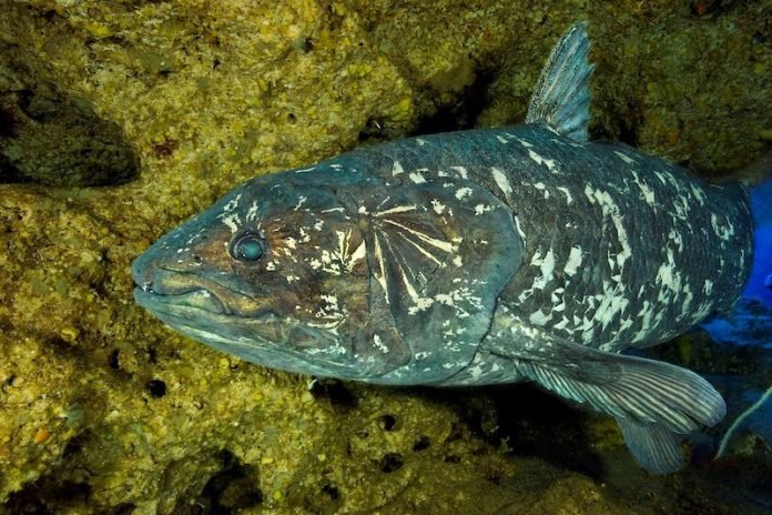
They studied one of the world’s oldest types of fish, Coelacanth.
The research was conducted by an international team led by the University of Bristol.
Coelacanth (Latimeria chalumnae) is very rare fish and it was thought to have gone extinct with dinosaurs over 65 million years ago.
Scientists discovered a living specimen in South Africa in 1938, and the finding led to a debate about whether this fish fits into the evolution of land animals.
Previous research has shown that the skull of this fish is completely split in half by special ‘intracranial joint’ and that its brain is very small.
This makes Coelacanth survival unique amongst all living vertebrates.
In the current study, the team studied the fish’s brain cavity at different stages of development to understand when the skull divides to form a hinged braincase.
The scientists used advanced imaging techniques to visualize the internal anatomy of the fish without damaging them.
And then they used the data to generate detailed 3-D models and analyzed how the form of the skull, the brain, and the notochord changes from a fetus to an adult.
They believe that the formation of this special joint may be caused by the unique development of the notochord (a tube extending below the brain and the spinal cord in the early stages of life).
It is very unusual especially compared to primates in which the brain expands dramatically.
The researchers also observed how these structures are positioned relative to each other at each stage.
They compared their observations with what is known about the formation of the skull in other vertebrates.
The team suggests that the findings provide new information about the unique skull of this fish and its links to the evolution of vertebrate species, including humans.
The lead author of the study is Dr. Hugo Dutel at the University of Bristol.
The study is published in Nature.
Copyright © 2019 Knowridge Science Report. All rights reserved.


